This thread has been locked.

If you have a related question, please click the "Ask a related question" button in the top right corner. The newly created question will be automatically linked to this question.

DLPNIRNANOEVM: different curves for same sample at different times, what is the reaon?

Part Number: DLPNIRNANOEVM

Hi,

I'm trying to do quantitative analysis for same liquid in a plastic cuvette(material:PS). However, when I scan the empty cuvette at afternoon and in the night, I found the curve is significantly differnt. Could you help to figure out what is the reason, and how to improve it?

The device I'm using is the default DLPNIRNANOEVM, with the reflectance scan head.

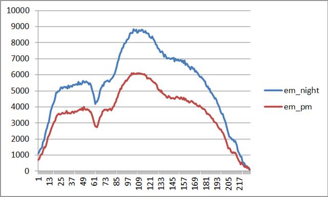
1.This is the sample signal for night & pm scan.

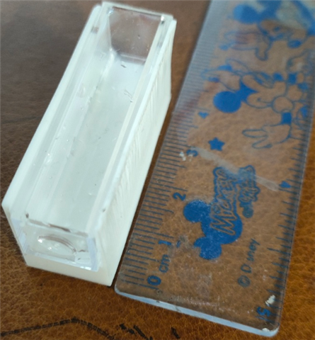
2.This is the sample, (no liquid inside, and I used a white plastic as background to enhance the reflect light).
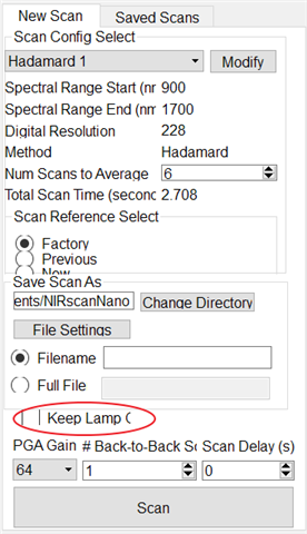
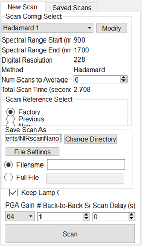
3.scan setup:

Above, thanks in advance!

Sichen Bian

  • Hi Sichen,

    Thanks for sharing the csv files. Upon analyzing the csv files, I noticed that the total scan time is different for both night scans are taking double the time to scan. 

    This could be possible if either the back to back or delay are changed.

    Please ensure that the back to back scans and scan delay are also set the same. 

    Regards,

    Akhil

  • Hi Akhil,

    The different scan time is because of lamp on/off: for ~2.6s scan, the lamp is off, for ~1.4s scan, the lamp is open.  I just tried to keep all the other same but change the "keep lamp on/off", and found a same curve as a result but different scan time.

    Some more csv might be helpful, i attached it. It is strange that,0604_night_lamp_off,0606_lamp_on/off are similar, but 0604_pm_lamp_on is very different. 0604_pm_lamp_on.csv0604_night_lamp_off.csv0606_lamp_off.csv0606_lamp_on.csv

    Sichen Bian

  • Hello Sichen,

    Thank you for this information. Please allow us time to look this over and investigate the behavior.

    Regards,

    Austin

  • Hello Sichen,

    There are Mutiple factors which could impact this:

     1. Sample presentation - any variation in sample presentation will cause change in absolute value of reflected /transmitted light. I assume you are using a transmissive head along with cuvette.  This system setup ensures consistent sample presentation.

    2. Hadamard pattern has a very high light output compared to column scan.  If you are scanning a blank cuvette and that too with PGA 64 , it is likely that the light out is too much and sensor is getting saturated:

      a. Please lower PGA gain to 1 and compare results.

      b. Use column scan and compare results.

    3. Lamp ON  flag - Keeping Lamp on between scans allows lamp to stay warm and stabilize light output for sequent scan. Your set up is averaging 6 scans.

      a. You can evaluate consistency of scan with "Lamp on" between scans vs "Lamp not on" between scans. Choose appropriate setting for you application.

     b. Change "Num scan to average" to 1 and see do you still see similar issue. You can optimize the number to average for time and consistency,

    Please go through these series of evaluations and select you configuration which offers consistent result for your use case.

    regards,

    Vivek